Whole-Body MRI Biomarkers Unlock A Fuller Picture In Neuromuscular Trials
By Weston Miller, MD, chief medical officer, Epicrispr

One of the challenges in early-stage clinical development for neuromuscular disease is knowing if a therapy is working soon enough and broadly enough to guide development decisions. Traditional functional endpoints such as timed walk tests, grip strength, or patient-reported outcomes are indispensable, but they are blunt tools. They often lag behind underlying biological changes and, perhaps more importantly, fail to capture the variability of disease progression across patients.
In neuromuscular disorders such as facioscapulohumeral muscular dystrophy (FSHD), some patients first experience weakness in their shoulders, while others notice it in their legs. If a trial endpoint focuses solely on gait, it risks missing a meaningful decline in upper body function. Conversely, an endpoint measuring arm strength could overlook deterioration in the lower body. The result is a fragmented view of disease, where the very signal of efficacy, or lack thereof, can be obscured by where the disease happens to strike first.
Beyond where the disease shows up, it also matters how much muscle is lost, even when that loss does not immediately affect a functional test. Skeletal muscle is not just an actuator for movement; it is a critical tissue with metabolic, structural, and hormonal roles. Losing it unnecessarily is never a good outcome.
Traditional Endpoints Mask Muscle Pathology That MRIs Can Reveal
The human musculoskeletal system has built-in redundancy. We have more muscles than are strictly necessary to perform most tasks. This means that when one muscle weakens or is injured, others can adjust and often take over its role. In some cases, synergist muscles grow even stronger to compensate. This redundancy allows people to maintain function even while individual muscles decline. But that does not mean the decline is unimportant. In fact, these kinds of changes can easily go unnoticed in standard outcome measures. Just as an athlete might still perform despite a muscle injury, patients may appear stable while their disease progresses at the muscle level.
Equally important is timing. Muscle pathology often begins months or even years before functional decline can be detected through traditional tests. This delay makes sense given the redundancy of the muscle system. When one muscle deteriorates, others can compensate — either by changing recruitment patterns through neural adaptation or by hypertrophying to take on more load. These strategies can preserve performance in the short term, but they also can mask early decline.
We shouldn’t penalize patients by relying only on metrics that miss the progression of disease simply because the body is still able to compensate. If anything, patients’ ability to adapt makes it even more important to use tools that can uncover what functional tests may miss. MRI-based biomarkers help reveal these hidden changes, accelerating the feedback loop between therapy and outcome. For trial sponsors and regulators, that means more confidence in early signals, more efficient trial designs, and potentially faster go/no-go decisions. In rare diseases, where recruiting patients is already a challenge, reducing the number of inconclusive or underpowered studies is critical.
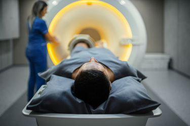
Whole-body MRI offers a way forward. It captures changes across all muscles, including those whose decline may be masked because other muscles are compensating for them or because they are not involved in the specific functional test being used. Quantitative measures such as muscle volume, fat infiltration, and inflammation can reveal early localized biological changes throughout the body. This reduces the risk of missing progression in “off-endpoint” muscle groups and creates a more complete, patient-centered picture of disease.
The Promise Of Whole-Body MRIs — For Sponsors, Patients, And Regulators
The technology has matured significantly. Advances in AI-driven segmentation and analysis now allow over 100 muscles throughout the body to be automatically quantified from a single scan. Epicrispr recently used Springbok Analytics’ AI-driven platform in our first-in-human clinical trial, and the partnership illustrates how emerging imaging analytics can be integrated into clinical trials to generate reproducible, regulator-ready data at scale. Workflows that once required hours of expert review can now be completed within minutes, and whole-body protocols can be executed in under 45 minutes. For sponsors, the availability of harmonized imaging data across global sites means results can be reproduced and compared across centers. For patients, MRI is noninvasive, widely available, and far less burdensome than invasive alternatives such as a muscle biopsy.
The implications extend beyond neuromuscular disease. Oncology, multiple sclerosis, and other areas have already demonstrated the value of imaging biomarkers where direct measures of function are limited. What is new is the convergence of imaging hardware, computational analytics, and standardized platforms capable of producing reproducible, regulator-ready data at scale. This convergence creates an opportunity to elevate imaging from a supportive exploratory tool to a decision-critical one.
Regulatory agencies are also signaling greater openness to advanced biomarkers. In other fields, imaging data have played roles as surrogate endpoints or validated biomarkers, helping accelerate drug approvals. In neuromuscular diseases where patient populations are small, progression is variable, and every datapoint carries disproportionate weight, the case for imaging is even stronger. The ability of MRI biomarkers to complement and contextualize functional outcomes makes it an increasingly important component of trial design.
That balance matters for patients. People with progressive neuromuscular conditions are less concerned with whether their improvement shows up first in a 6-minute walk test or a grip strength measurement. They want assurance that a therapy is slowing, stopping, or reversing muscle deterioration wherever it appears. Whole-body imaging offers a way to honor that lived reality. By capturing the totality of disease burden, it ensures that progress or decline is not hidden simply because a muscle group was outside the scope of a trial endpoint.
Considerations For The Use Of Whole-Body MRIs As A Digital Biomarker
There are practical considerations as well. Imaging introduces new costs, workflows, and analytical requirements. Sponsors must weigh these against the value of more sensitive and comprehensive data. Yet as automation and AI reduce time and cost, the calculus is shifting. The choice may no longer be between traditional endpoints and imaging endpoints but rather how best to integrate them in a complementary way that strengthens both.
Of course, enthusiasm should be tempered by caution. Imaging data must be collected and analyzed under rigorous quality management systems to avoid variability across sites. Large-scale validation is needed before imaging biomarkers can routinely serve as surrogate endpoints. But progress is clear. Pilot programs are moving imaging from the exploratory margins toward the regulatory mainstream, creating a path that others can follow and refine.
Whole-body MRI is also beginning to reveal new insights about disease biology itself. In FSHD, for example, a consistent spatial pattern of muscle involvement has emerged from a recently published multiscale model that combines MRI and clinical data to predict where and how fast each muscle (and region within a muscle) will decline. That model builds a “digital twin” for each patient to simulate untreated disease progression, which could serve as a surrogate placebo in trials. These kinds of models suggest that imaging could play a valuable role not just in measuring disease but in predicting it, helping trials detect efficacy even when functional decline is masked by compensation or when endpoints don’t include all affected muscles.
What’s Ahead For Whole-Body MRIs As Biomarkers
For the clinical trial community, the challenge now is not whether to use whole-body imaging but how to incorporate it wisely and consistently. Should it be built into early-phase studies to identify promising therapies faster? Should it become a secondary endpoint in pivotal trials to complement functional outcomes? Should it be standardized across sponsors to enable cross-trial comparisons? And as predictive models improve, should we begin using digital twins — personalized projections of untreated disease — to simulate control arms or serve as surrogate placebos? These are the questions we must begin to answer collectively.
For a field where every patient enrolled is precious, and every data point carries disproportionate weight, whole-body imaging is more than an incremental improvement. It represents a shift in how we define and detect efficacy. As clinical trials move toward increasingly precise patient-centered evaluation, the ability to see all of the disease — wherever it manifests — may be one of the most important advances we can make, because every muscle matters.
About The Author:
 Weston (Wes) Miller, MD, is the chief medical officer at Epicrispr. Dr. Miller has experience in the clinical development of in vivo and ex vivo genomic medicines for patients with rare severe genetic disorders.
He began his career as an associate professor of pediatrics in the division of pediatric blood and marrow transplantation (BMT) at the University of Minnesota. At Sangamo Therapeutics, he led programs investigating ex vivo disruptive gene editing of autologous hematopoietic stem and progenitor cells for patients with severe beta globinopathies, as well as in vivo gene editing for patients with mucopolysaccharidosis types I and II. At Astellas Gene Therapies, he and his team led programs investigating in vivo gene transfer for patients with X-linked myotubular myopathy and Pompe disease. At Graphite Bio, he led the investigation of an ex vivo gene-corrected autologous hematopoietic stem cell product for patients with severe sickle cell disease.
Dr. Miller received his B.S. in chemistry from Stanford University and his MD from Louisiana State University School of Medicine, New Orleans.
