
Why Radioligand Therapy Studies Look Nothing Like Standard Oncology Phase 1s
A conversation between Affibody Chief Scientific Officer Fredrik Frejd and Clinical Leader Executive Editor Abby Proch
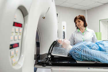
Radiopharmaceutical trials demand quite a bit more operational and logistics expertise beyond that of a standard oncology study. In this interview, Affibody CSO Fredrik Frejd explains why radioligand therapy (RLT) development depends on specialized sites, nuclear medicine expertise, imaging capabilities, and rigorous dosimetry. He explores how radiation deposited in healthy organs shapes safety assessments and dose-escalation decisions, while delayed toxicities complicate traditional monitoring. Frejd also examines the trade-offs between individualized dosing and fixed-dose approaches, as well as the need for sustained regulatory support in the U.S. and beyond.
Clinical Leader: When radiopharmaceutical trials are run at sites without nuclear medicine infrastructure and radiation‑certified personnel, what specific risks or failure points might emerge?
Fredrik Frejd: A very important part of running first-in-human and early trials is understanding the distribution of the radioactivity. This is called dosimetry, which is how much energy is deposited in different organs. From that, you can calculate the risk to certain organs, which means that you can understand safety for the patient.
It can differ depending on what type of agent you administer. Small peptides will most likely have a high kidney dose. Large molecules, such as antibodies, will likely have a substantial amount of exposure in the blood. Small molecules can be found anywhere.
Toxicity for radioactivity is often focused on slow and accumulating late effects, not the classical toxicity you see early on. This means that if you administer too much to the kidney, you might only discover the toxic effect years later when the kidneys stop functioning.
Therefore, you need to measure radioactivity distribution and monitor for late toxicity events early on. This is why early-phase trials must be done in facilities with imaging capabilities. You can then quantify the amount of deposited energy in the different organs, analyze it, and then determine safe dose escalation. That's a lot of expense and difficulty.
There is also an ongoing discussion between nuclear medicine and oncology. Some argue for more classical dose-escalation studies that monitor for, safety, PK and toxicity. Others want to focus more on the dosimetry calculations to tailor the administration of radioactivity to each patient.
For example, for a physician to administer a tailored amount of radioactivity, it depends on the weight and the physiology of the patient. Ideally, the physician would first conduct a diagnostic scan with a tracer or agent that is to be delivered with a small amount of radioactivity or a diagnostic surrogate molecule. That way, he or she can understand perfectly how it all distributes in different organs and can then calculate exactly this patient’s ideal dose.
In contrast are the latest radiopharmaceutical guidelines from the FDA, which lean towards one fixed dose on a defined schedule that is safe for everyone. However, that likely means that some patients could have tolerated a higher dose and perhaps achieved a higher clinical benefit but are instead underdosed.
How do controlled facilities and trained staff directly influence patient safety, regulatory compliance, and the reliability of trial data?
Let's make a hypothetical example. You, as a patient, receive a standard drug. You go to an infusion center, get your standardized dose, and then off you go with the normal safety checks as follow-up. It's a very easy and accessible dosing regimen.
The opposite is that you need to go to a university hospital. There, they have imaging equipment and trained staff, everything. They do the diagnostic scan, calculate your exact dose, administer the dose, and then repeat as needed.
That is much more controlled, and you get much more reliable data. But it will be less accessible to a wider patient population and negatively impact the trial recruitment rate. So, it's a delicate balance of robustness of data versus speed of the trial and, hence, development.
The regulatory bodies note that the latter isn’t available to a broad mass of patients. So, to make it available to broad populations, the challenge is to have enough rigor to find a safe recommendation in their guidance.

What additional role does dosimetry play, alongside PK/PD and toxicity, in defining safe and effective dose-escalation pathways for radiotherapeutics?
Information from dosimetry adds information on how much energy is deposited in different organs, and that translates to safety. It will tell quite a lot about how different organs react.
You can predict that if you double the radioactive dose and have the same type and amount of trace carrier vehicle of the radioactivity in the tissue, it's scalable. You can start with a low radioactive dose and get an understanding of how a high radioactive dose would behave, including the maximum dose.
On the other hand, it is very difficult to predict effects of microdosimetry, which is done at the cellular level of very small structures. For example, even a small amount of target expression in the bone marrow could cause certain cells to take up the radiopharmaceutical and receive a higher radiation dose than we are able to detect through conventional imaging. This means the desired toxicity could occur at a lower administered activity than the overall dosimetry would predict.
Often in practical clinical trials, the escalation decisions are based more on safety. That is, we have not yet reached too high a dose to different organs, and therefore we can proceed, rather than using dosimetry only to draw this conclusion.
But in principle, one could use it both ways. Dosimetry could guide the escalation decisions to make them much more precise and make both bigger steps or smaller steps, depending on where in the escalation curve you are.
What unique insights do nuclear medicine physicians, dosimetrists, and radiation safety officers contribute that traditional early‑phase review committees cannot replicate?
It's absolutely crucial to have multidisciplinary trial review committees. Without a hospital physicist, a nuclear medicine physician, and probably a radiologist it would be impossible for the oncologist to understand the risk.
It could also be challenging for the physicists to understand what tumor lesions to measure or where the tumors are located without the nuclear medicine physician and the radiologist. The team will set the different limits for different organs and uptake in tumors, which guides the benefit-risk assessment. An oncologist cannot master these techniques.
It's crucial that each comes together to form a broad review, looking not only at blood chemistry and safety signals but also calculating the radioactive doses to different organs so they can establish a safe dose.
Without having information about the dosimetry, it would be very risky to do escalations because you don't really know where the activity accumulates and exerts its toxicity.
How could radiopharmaceuticals trials be improved?
Some say using minimal amounts of carrier, sub-toxic amounts, or sub-pharmacological amounts would allow for early imaging and, therefore, a better understanding of its behavior.
In research and early development, it's absolutely crucial to access early data to understand the distribution of the drug. Radiolabeled tracers can be detected with very high sensitivity, so you can get a lot of information even with minute amounts of tracer or targeting agent.
Ideally, this could happen before making the massive investment in final manufacturing of the drug and full process validation of the radiolabeling. Otherwise, you can incur significant costs before getting the first few images that might actually tell you that the drug will not work.
Now, the frameworks being developed in Europe and the U.S. are treating radiopharmaceuticals more like conventional drug development (and introducing time-consuming requirements), where previous tradition more often could utilize a microdosing-like procedure with a slimmer path to first-in-human images. That can make it less efficient to translate promising radiopharmaceutical research into clinical development. A wish would be to find a path where we could do this in a safe and controlled fashion in both locations.
It's fine to impose the requirements for a later-stage project, but at the early stage, where early data influences further investment, it’s not feasible, and we need to have an educated, scientifically based discussion with agencies about this.
About The Expert:
 Fredrik Frejd is the chief scientific officer at Affibody and has over 25 years of experience in life science research with particular expertise in biologics drug development, phage display, and therapeutic protein engineering of alternative scaffolds. Dr. Frejd received his Ph.D. from the Swiss Federal Institute of Technology, ETH, in Zurich in 2001 and joined Affibody AB in 2002. Dr. Frejd is also an adjunct professor in cancer precision medicine at the Department of Immunology, Genetics and Pathology at Uppsala University, with a special focus on the development of tumor-targeting agents for molecular radiotherapy.